File size: 5,215 Bytes
139850b 32f3b11 139850b ef9db36 139850b 6ee0257 139850b ef4cf37 139850b ef9db36 139850b 32f3b11 |
1 2 3 4 5 6 7 8 9 10 11 12 13 14 15 16 17 18 19 20 21 22 23 24 25 26 27 28 29 30 31 32 33 34 35 36 37 38 39 40 41 42 43 44 45 46 47 48 49 50 51 52 53 54 55 56 57 58 59 60 61 62 63 64 65 66 67 68 69 70 71 72 73 74 75 76 77 78 79 80 81 82 83 84 85 86 87 88 89 90 91 92 93 94 95 96 97 98 99 100 101 102 103 104 105 106 107 108 109 110 111 112 113 114 115 116 117 118 119 120 121 122 123 124 125 126 127 128 129 130 131 132 133 |
---
tags:
- monai
- medical
library_name: monai
license: apache-2.0
---
# Model Overview
A pre-trained model for volumetric (3D) segmentation of brain tumor subregions from multimodal MRIs based on BraTS 2018 data. The whole pipeline is modified from [clara_pt_brain_mri_segmentation](https://catalog.ngc.nvidia.com/orgs/nvidia/teams/med/models/clara_pt_brain_mri_segmentation).
## Workflow
The model is trained to segment 3 nested subregions of primary brain tumors (gliomas): the "enhancing tumor" (ET), the "tumor core" (TC), the "whole tumor" (WT) based on 4 aligned input MRI scans (T1c, T1, T2, FLAIR).
- The ET is described by areas that show hyper intensity in T1c when compared to T1, but also when compared to "healthy" white matter in T1c.
- The TC describes the bulk of the tumor, which is what is typically resected. The TC entails the ET, as well as the necrotic (fluid-filled) and the non-enhancing (solid) parts of the tumor.
- The WT describes the complete extent of the disease, as it entails the TC and the peritumoral edema (ED), which is typically depicted by hyper-intense signal in FLAIR.

## Data
The training data is from the [Multimodal Brain Tumor Segmentation Challenge (BraTS) 2018](https://www.med.upenn.edu/cbica/sbia/brats2018/tasks.html).
- Target: 3 tumor subregions
- Task: Segmentation
- Modality: MRI
- Size: 285 3D volumes (4 channels each)
The provided labelled data was partitioned, based on our own split, into training (200 studies), validation (42 studies) and testing (43 studies) datasets.
Please run `scripts/prepare_datalist.py` to produce the data list. The command is like:
```
python scripts/prepare_datalist.py --path your-brats18-dataset-path
```
## Training configuration
This model utilized a similar approach described in 3D MRI brain tumor segmentation
using autoencoder regularization, which was a winning method in BraTS2018 [1]. The training was performed with the following:
- GPU: At least 16GB of GPU memory.
- Actual Model Input: 224 x 224 x 144
- AMP: True
- Optimizer: Adam
- Learning Rate: 1e-4
- Loss: DiceLoss
## Input
Input: 4 channel MRI (4 aligned MRIs T1c, T1, T2, FLAIR at 1x1x1 mm)
1. Normalizing to unit std with zero mean
2. Randomly cropping to (224, 224, 144)
3. Randomly spatial flipping
4. Randomly scaling and shifting intensity of the volume
## Output
Output: 3 channels
- Label 0: TC tumor subregion
- Label 1: WT tumor subregion
- Label 2: ET tumor subregion
## Model Performance
The achieved Dice scores on the validation data are:
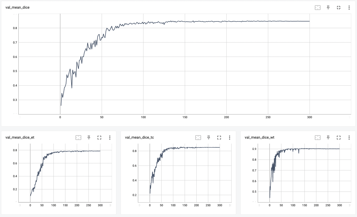
- Tumor core (TC): 0.8559
- Whole tumor (WT): 0.9026
- Enhancing tumor (ET): 0.7905
- Average: 0.8518
## commands example
Execute training:
```
python -m monai.bundle run training --meta_file configs/metadata.json --config_file configs/train.json --logging_file configs/logging.conf
```
Override the `train` config to execute multi-GPU training:
```
torchrun --standalone --nnodes=1 --nproc_per_node=8 -m monai.bundle run training --meta_file configs/metadata.json --config_file "['configs/train.json','configs/multi_gpu_train.json']" --logging_file configs/logging.conf
```
Please note that the distributed training related options depend on the actual running environment, thus you may need to remove `--standalone`, modify `--nnodes` or do some other necessary changes according to the machine you used.
Please refer to [pytorch's official tutorial](https://pytorch.org/tutorials/intermediate/ddp_tutorial.html) for more details.
Override the `train` config to execute evaluation with the trained model:
```
python -m monai.bundle run evaluating --meta_file configs/metadata.json --config_file "['configs/train.json','configs/evaluate.json']" --logging_file configs/logging.conf
```
Execute inference:
```
python -m monai.bundle run evaluating --meta_file configs/metadata.json --config_file configs/inference.json --logging_file configs/logging.conf
```
# Training
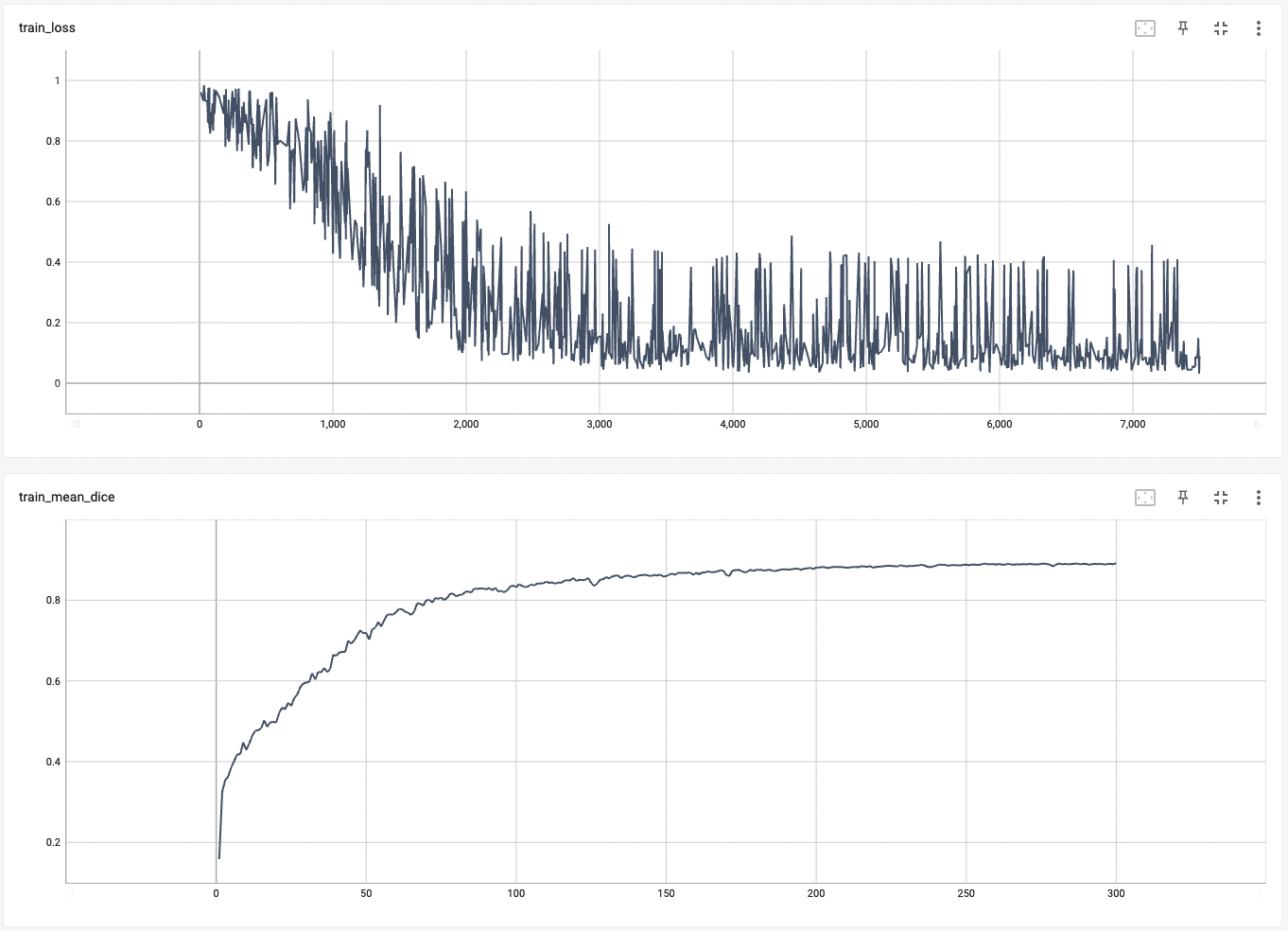
A graph showing the training loss and the mean dice over 300 epochs.

# Validation
A graph showing the validation mean dice over 300 epochs.

# Disclaimer
This is an example, not to be used for diagnostic purposes.
# References
[1] Myronenko, Andriy. "3D MRI brain tumor segmentation using autoencoder regularization." International MICCAI Brainlesion Workshop. Springer, Cham, 2018. https://arxiv.org/abs/1810.11654.
# License
Copyright (c) MONAI Consortium
Licensed under the Apache License, Version 2.0 (the "License");
you may not use this file except in compliance with the License.
You may obtain a copy of the License at
http://www.apache.org/licenses/LICENSE-2.0
Unless required by applicable law or agreed to in writing, software
distributed under the License is distributed on an "AS IS" BASIS,
WITHOUT WARRANTIES OR CONDITIONS OF ANY KIND, either express or implied.
See the License for the specific language governing permissions and
limitations under the License.
|